
MedClinics News & Blog
Spina bifida treatment is usually discussed at the moment of diagnosis, when parents are already being asked to understand anatomy, surgery, timing, risk, and the long-term needs of a child they have not yet met.
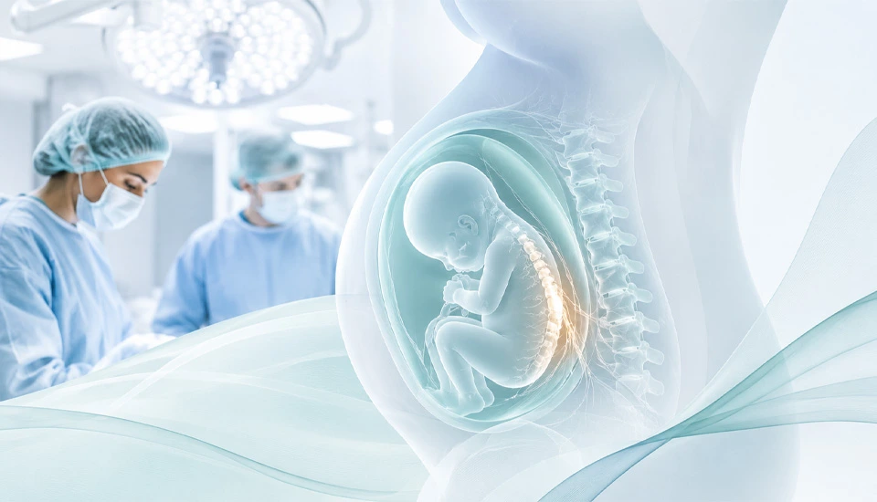
But in the most severe form, myelomeningocele, the condition has already been unfolding for some time. Part of the baby’s spinal cord and surrounding nerves remain exposed through an opening in the spine. That exposure matters because the nerve tissue is developing while it is also vulnerable.
This is why fetal surgery became such an important part of the story. Repairing the opening before birth can protect the spinal cord earlier than surgery after delivery. It changed what was possible for selected cases, even though it also brought the complexity of operating before birth, with all the care that requires for both mother and baby.
Now researchers at UC Davis have added another layer to that conversation.
In a Phase 1 study published in The Lancet, the team tested a small patch containing placenta-derived mesenchymal stem cells during fetal repair of myelomeningocele. The patch was placed directly over the exposed spinal cord area during surgery. The first group was small – six babies – and the early focus was safety. The patch could be placed during the procedure, and UC Davis reported no early stem-cell-related tumors, infections, spinal fluid leaks, or abnormal tissue growth in that first treated group.
For fetal medicine, that kind of early result carries weight. Before the field can ask whether an added therapy changes long-term function, it first has to know whether the approach can be integrated into an already demanding operation.
That is what makes this study worth reading carefully.
Table of Contents
Why This Matters for Spina Bifida Treatment
The idea behind fetal repair is not new. It developed from a simple but difficult observation: in myelomeningocele, some of the damage happens before birth.
For this reason, the timing of spina bifida treatment has become an important part of fetal medicine.
For many years, surgery was performed after delivery. The defect could be closed, and the newborn could begin care. But the spinal cord had already spent months exposed during pregnancy. Fetal surgery shifted the timing earlier.
The landmark MOMS trial helped establish the value of that shift. Prenatal repair reduced the need for shunting and improved some motor outcomes in selected babies compared with postnatal repair. It became one of the clearest examples of fetal surgery changing the course of a condition before birth.
Even so, fetal repair has never been a simple endpoint. The procedure is highly specialized. It requires careful selection and experienced teams. Many children still need years of neurological, orthopedic, urological, and rehabilitation care. The surgery can protect the spinal cord earlier, but myelomeningocele remains a complex developmental condition.
That is the background for the UC Davis trial. It is not starting from the idea that closure alone has solved the problem. It is asking whether the repair site can be given more biological support at the same moment the defect is closed.
What the Stem Cell Patch Adds

The treatment used in the study was precise.
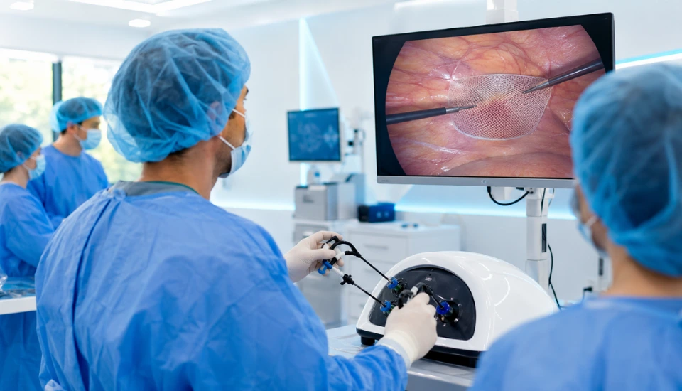
The cells were derived from placental tissue and placed on a biological scaffold. During fetal surgery, the patch was applied locally over the exposed spinal cord area before the repair was completed.
That local placement is the heart of the study.
Mesenchymal stem cells are often studied less for what they become and more for what they release. They can produce signals that may influence inflammation, tissue protection, and healing. In this setting, the interest is in whether those signals can improve the environment around vulnerable nerve tissue while development is still underway.
This is a more grounded way to think about stem cell therapy. The cells are not being treated as a broad answer to spina bifida. They are being used as part of a specific surgical moment, in a specific location, where the biology of protection and repair may matter.
That is also why the fetal setting is so important. The exposed spinal cord is not an old injury. It is part of an active developmental process. The tissue is still forming, still changing, and still being shaped by the environment around it.
That timing gives the study its logic.
What the First Results Show
The first six cases mainly tell us that the approach could be carried out during fetal repair and that the early safety profile was reassuring enough for the study to continue.
The more practical questions will take longer.
Movement is not fully understood at birth. Bladder and bowel function develop over time. Independence, mobility, the need for braces or other supports, and the child’s day-to-day function all become clearer only as children grow.
UC Davis has said the children will be followed through early childhood. That follow-up will matter more than any first report, because the value of this approach will depend on whether the early safety signal is followed by meaningful functional outcomes.
That is often how serious regenerative medicine develops. The early study answers a narrow question. Later follow-up decides whether the biological idea has clinical weight.
Why This Matters for Regenerative Medicine

Stem cell medicine has often suffered from language that is too broad. The word “regeneration” can make a study sound more complete than it is. It can also make very different approaches seem as if they belong to one simple category.
This trial is useful because it is much more specific than that.
It is about one cell source, used in one surgical setting, at one moment in fetal development. The goal is not to present stem cells as a general treatment, but to see whether they can support a repair that surgeons are already performing.
That specificity is where the field becomes more serious.
Regenerative medicine is gradually moving away from the idea that repair is simply a matter of adding cells. More of the discussion now is about signals, timing, tissue environment, immune response, and how to guide healing in a controlled way. Stem cells are part of that conversation. So are exosomes, engineered tissues, gene editing, and small molecules that change how cells behave.
The UC Davis study belongs to that broader shift. It treats regeneration less as a slogan and more as a practical question: can the right biological support, delivered at the right time, improve the outcome of an existing medical procedure?
In fetal spina bifida repair, that question is especially powerful because timing is already central to the treatment.
A Different Way to Think About Repair Before Birth
The most interesting part of the study may be the idea that fetal surgery could become more than structural repair.
For years, the main aim has been to close the spinal opening early enough to protect the cord from further exposure. That remains essential. But the stem cell patch adds another possibility: the repair site itself may become biologically active, offering local support to tissue that is still developing.
That is a subtle shift, but an important one.
If longer follow-up shows stronger motor outcomes, better bladder or bowel function, or fewer long-term complications, this early study may become part of a larger change in how myelomeningocele is approached. If the benefits are more limited, it will still have helped define how cell-based therapies can be tested responsibly in fetal surgery.
Either way, it moves the conversation forward.
The study is small, but the question behind it is not. It asks whether regenerative medicine can be used in one of the most time-sensitive areas of care, not as a separate treatment, but as part of a surgical repair performed before birth.
For a condition where early development can shape lifelong function, that is a serious question to follow.
Frequently Asked Questions About Spina Bifida Treatment Before Birth
What makes this study different from standard fetal surgery?
Standard fetal surgery for myelomeningocele focuses on closing the spinal opening before birth. In this study, the UC Davis team added something extra to that repair: a small patch carrying placenta-derived mesenchymal stem cells. The patch was placed directly over the exposed spinal cord area during the operation.
Why use stem cells in this setting?
The interest is not only in closing the defect, but in supporting the tissue around it. In myelomeningocele, the spinal cord is exposed while the baby is still developing. Researchers are studying whether stem cells placed at the repair site may help create a more protective healing environment during that early window.
Are these stem cells meant to become new spinal cord tissue?
That is not the main idea. Placenta-derived mesenchymal stem cells are mainly being studied for the signals they release. These signals may influence inflammation, tissue protection and repair. In this trial, the cells were used as local biological support during surgery.
What did the first treated cases show?
In the first six babies, the stem cell patch could be placed during fetal repair, and the early safety findings were reassuring. UC Davis reported no early stem-cell-related tumors, infections, spinal fluid leaks or abnormal tissue growth in this first group.
What still needs to be followed over time?
The most important questions will come as the children grow. Researchers will be looking at movement, bladder and bowel function, daily development, and long-term safety. These outcomes cannot be judged fully at birth.
Is this available for patients now?
This approach is still part of a clinical trial. It is not standard care, and fetal surgery itself is only considered in carefully selected cases at specialized centers.
Why is this important for regenerative medicine?
It shows a more precise direction for stem cell research. The cells are not being used as a general treatment, but as a targeted addition to fetal surgery, at a moment when the developing spinal cord may still be influenced by its local environment.
Sources
- Farmer DL, Wang A, et al. Feasibility and safety of cellular therapy for in-utero repair of myelomeningocele: CuRe Trial. The Lancet. 2026.
- UC Davis Health. First-ever in-utero stem cell therapy for fetal spina bifida repair is safe, study finds. 2026.
- ClinicalTrials.gov. Cellular Therapy for In Utero Repair of Myelomeningocele – The CuRe Trial.
- ACOG / SMFM. Maternal-Fetal Surgery for Myelomeningocele.