Clinical Insights from Prof. Dr. Serdar Kabataş, MD, PhD (C)
MedClinics News & Blog
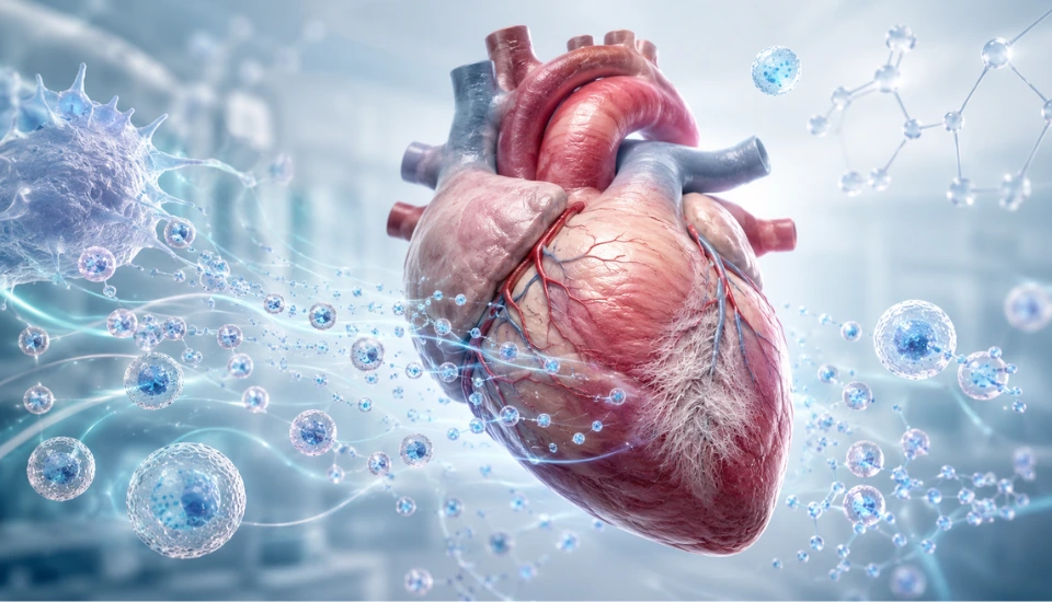
MSC extracellular vesicles after heart attack are being studied for a very specific reason: the damage is not over when blood flow returns.
That is the part many people do not see.
In a heart attack, the urgent problem is the blocked artery. The artery has to be opened quickly. Blood flow has to come back. The aim is to save as much heart muscle as possible before the injury becomes permanent.
That first step is essential.
But the heart still has work to do afterward.
The injured area has to repair itself. Immune cells arrive. Fibroblasts become active. The tissue starts laying down scar. Some of this is necessary. A damaged area needs structure. The heart cannot leave the injured wall weak.
But too much scar has a cost.
Heart muscle has to move. It has to fill, contract and push blood forward. Scar tissue does not do that job like healthy myocardium. If fibrosis becomes too active after myocardial infarction, the ventricle can become stiffer and less efficient over time.
This is the problem a new Cell Stem Cell study looks at.
The paper was published online on July 24, 2026. Researchers studied an extracellular vesicle-enriched secretome derived from bone marrow mesenchymal stromal cells, or MSCs. The product was tested in cell experiments, mouse models and a pig model of myocardial ischemia-reperfusion injury.
No patient received this MSC-derived product as treatment. The human part of the paper was related to imaging fibrotic activity after STEMI.
That is important, but it should not make the study feel small. Good preclinical work can still be valuable when the question is precise. Here, the question is not whether “stem cells repair the heart” in a general way. The question is whether MSC-derived signals can reduce the fibrotic response that follows ischemic heart injury.
That is a much more useful place to begin.
Table of Contents
The Problem After the Heart Attack
A heart attack is not only an event. It is followed by a process.
Once blood flow is restored, the heart enters a repair phase. The tissue has already been stressed by oxygen loss. Some cells are damaged or dead. The immune system responds. Fibroblasts begin to reshape the local environment.
At first, fibrosis is not the enemy.
Scar formation helps stabilize injured tissue. Without it, the damaged region would be fragile. In that sense, fibrosis is part of healing.
The difficulty is balance.
Too little structural repair would be dangerous. Too much fibrosis can also become a problem. The left ventricle may lose flexibility. Pumping function may decline. Over months, this kind of remodeling can contribute to heart failure after myocardial infarction.
That is why cardiac fibrosis has become an important target in post-heart attack research.
It is not as dramatic as the moment the artery is reopened. It is slower. It is less visible. But it may shape what kind of recovery the patient has later.
What MSC Extracellular Vesicles Are
The product in the study came from bone marrow MSCs. MSC stands for mesenchymal stromal cells, a term often used in scientific literature for this type of cell source.
MSCs are often discussed under the wider stem cell umbrella. In this study, the interesting part is not that the cells were transplanted into the heart. They were not. The researchers looked at what the MSCs released.
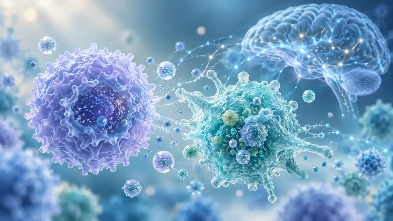
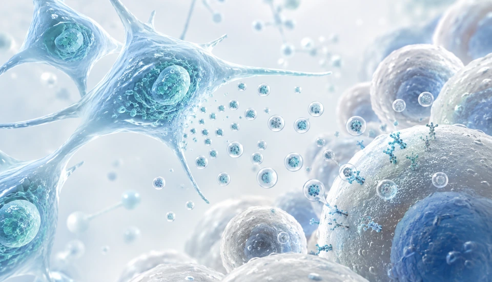
Cells release signals into their surroundings. MSCs can release proteins, lipids, RNA-containing particles and extracellular vesicles. Together, this released material is often called the secretome.
Extracellular vesicles, or EVs, are small membrane-bound particles. They can carry biological messages from one cell to another. Exosomes are one type of extracellular vesicle, but EV is the broader term.
The study used an EV-enriched secretome. That means the preparation contained extracellular vesicles and other MSC-secreted factors. It was cell-free, but it still came from MSC biology.
This is one direction regenerative medicine is taking: not only asking what cells can become, but also what signals they send.
For the heart, that matters.
Repair after myocardial infarction involves more than heart muscle cells. Fibroblasts, immune cells, endothelial cells and extracellular matrix all take part. A secretome-based product may influence the repair environment in more than one way.
That is the biological logic behind the study.
Why Laminin-521 Matters
One detail in the paper is easy to overlook, but it changes the story.
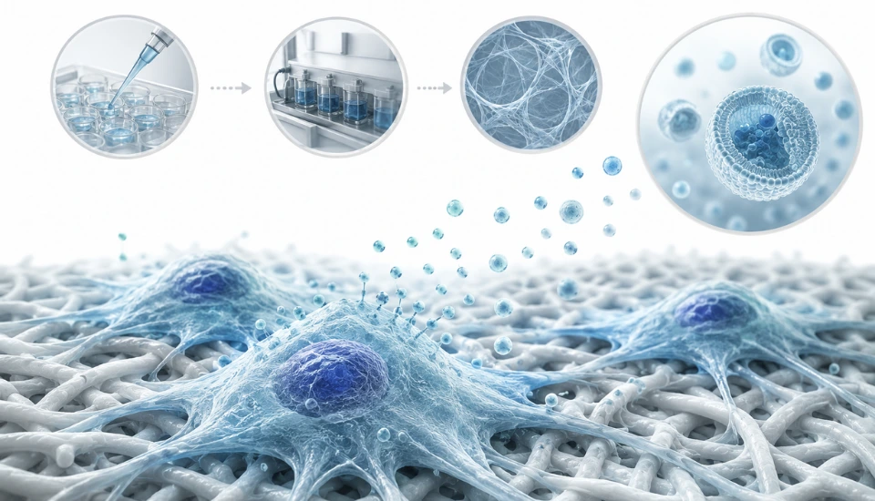
The researchers did not grow all MSCs in the same way. They compared different culture conditions. One method used laminin-521, a defined extracellular matrix protein.
MSCs grown with laminin-521 released an EV-enriched secretome with stronger anti-fibrotic activity than secretome produced under standard culture conditions.
This is not just a lab detail.
Cells respond to the surface they grow on. They respond to the matrix around them. The culture environment can change what they release. With extracellular vesicles, that can mean changes in the cargo, the activity and the final biological effect.
So the study is not only about MSC-derived extracellular vesicles. It is also about how the product is made.
That point is practical. If a cell-free MSC-derived product is ever going to be tested clinically, it has to be produced in a controlled way. The product has to be consistent. The manufacturing process has to be reproducible. Researchers need to know that one batch is behaving like the next.
In this paper, laminin-521 is part of that direction. It suggests that the culture environment may help shape an MSC secretome toward a more anti-fibrotic profile.
What Happened in the Models
The study moved step by step.
First, the researchers looked at fibrosis-related activity in cell-based experiments. Then they tested the EV-enriched secretome in mouse models of myocardial ischemia-reperfusion injury.
In the mouse models, the product was linked with less cardiac fibrosis and better preservation of left ventricular function. The paper also reported lower activation of PDGFRβ-associated myofibroblasts.
Myofibroblasts are important here because they are closely involved in scar formation. When they remain highly active, the tissue can become more fibrotic.
The study also described changes in macrophage behavior. Macrophages are immune cells, but they are also part of tissue repair. Depending on their state, they can support inflammation or help move the tissue toward repair. In this study, the MSC-derived secretome was associated with a more reparative macrophage profile.
That fits the wider idea of MSC signaling. The effect is not only about one cell type. It is about the repair environment.
The pig model added another layer.
Pig hearts are closer to human hearts than mouse hearts in size and physiology. That does not make them the same as human hearts, but it does make them useful in cardiovascular translational research.
In the pig model, the product was delivered through the coronary circulation after ischemia-reperfusion injury. The researchers reported cardioprotective effects.
This still does not answer the human question. It does make the preclinical signal stronger than a mouse-only study.
Why This Is a Fibrosis Story
The word “regeneration” can become too broad.
In the heart, it can also create the wrong picture. After myocardial infarction, the adult human heart does not simply rebuild lost muscle in a complete way. The repair process depends heavily on scar formation.
That is why this study is better understood as a fibrosis story.
The aim is not to erase healing. The aim is to prevent repair from becoming too damaging.
If fibrosis can be reduced in the right way, the ventricle may be protected from some of the stiffness and remodeling that can follow a heart attack. That would be meaningful even if the treatment does not replace every lost cardiomyocyte.
This is where MSC-derived extracellular vesicles after heart attack become interesting.
Their possible role is not only “regeneration” in the dramatic sense. It is signaling. It is immune modulation. It is fibroblast behavior. It is how the injured tissue decides what kind of scar to build.
That is a quieter kind of regenerative medicine.
But it may be closer to the way some therapies actually move forward.
The Imaging Part
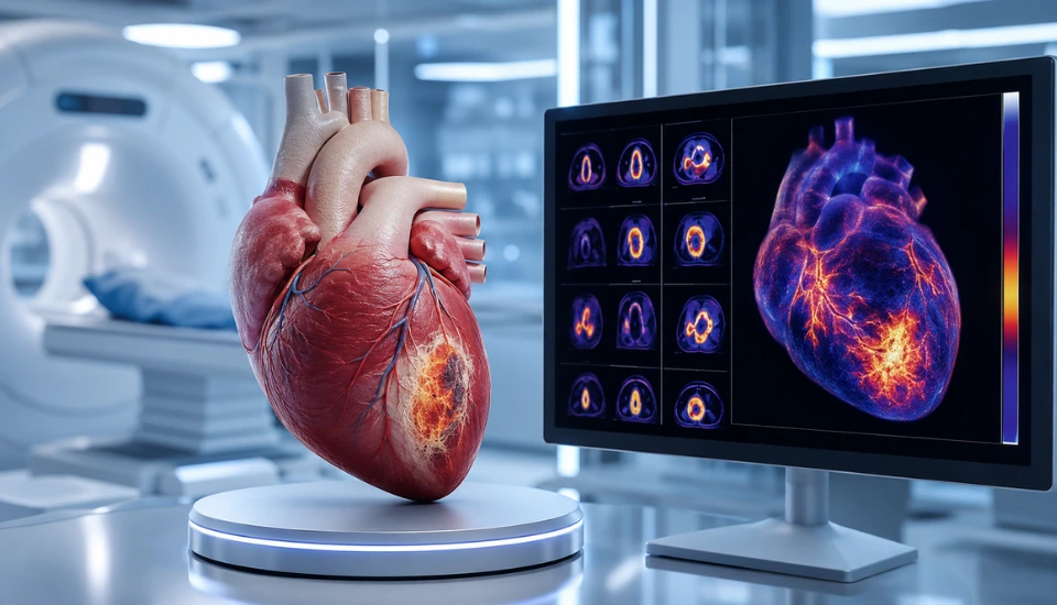
The paper also included PDGFRβ-targeted PET imaging.
This was used to look at fibrotic activity and myofibroblast activation. The researchers also described imaging observations in STEMI patients, suggesting that myofibroblast activity may continue for weeks after the acute heart attack.
Those patients were not treated with the MSC-derived EV-enriched secretome.
The imaging part is still useful because it points to a future clinical problem. If researchers want to target fibrosis after myocardial infarction, they need to know when that fibrosis is active. They also need a way to follow whether an intervention is changing the biology it is supposed to change.
Timing matters.
A treatment aimed at fibrosis would not be like a general supplement given at any point. It would need a window. It would need patient selection. It would need a measurable target.
Imaging may help define that in future studies.
Why This Study Deserves Attention
This study is not a new treatment for heart attack patients. It is not proof that extracellular vesicles prevent heart failure in humans. It is also not “just” another broad “stem cell” story.
Its value is more specific.
The researchers worked with a defined MSC-derived EV-enriched secretome. They connected the product to laminin-521 culture conditions. They studied cardiac fibrosis after ischemia-reperfusion injury. They used cell systems, mouse models and a pig model. They also included imaging work related to fibrotic activity in STEMI patients.
That is why the paper feels relevant for regenerative medicine.
The field is moving away from general language and toward defined biological roles. A product is not important only because it comes from MSCs or contains extracellular vesicles. It becomes important when the product, target, model and possible clinical pathway are clear.
Here, the target is post-ischemic cardiac fibrosis.
Heart failure after myocardial infarction remains difficult, even with modern acute care. If a future therapy could safely reduce harmful remodeling after the acute event, it would matter.
This study does not take the field there yet. It gives the field a reason to keep studying that direction.
What Comes Next
Several questions remain.
When after myocardial infarction would this type of product make sense? What dose would be safe? Would intracoronary delivery be practical in patients? Which patients would have enough active fibrosis to be considered? How should response be measured? Could PET imaging help select or monitor patients?
These are not small details. They are the clinical work that would have to come next.
The manufacturing side also matters. EV-enriched secretome products need clear quality control. The culture method, vesicle content, potency testing and batch consistency would all be part of any serious development pathway.
That is why the laminin-521 part of the study is worth noticing. It suggests that product design may shape biological effect.
For now, the paper adds a strong preclinical signal to regenerative cardiology. It shows that MSC-derived extracellular vesicles after heart attack may be studied not as a vague repair idea, but as a possible way to influence fibrosis and remodeling after ischemic injury.
That is enough to make the study worth following.
Frequently Asked Questions About MSC-Derived Extracellular Vesicles After Heart Attack
What are MSC-derived extracellular vesicles?
MSC-derived extracellular vesicles are tiny particles released by mesenchymal stromal cells. They can carry biological signals such as proteins, lipids and RNA. Researchers study them because MSCs may affect injured tissue partly through the signals they release.
What did the Cell Stem Cell study investigate?
The study investigated an extracellular vesicle-enriched secretome from bone marrow MSCs. Researchers tested whether this cell-free product could reduce fibrosis and help preserve heart function after ischemic heart injury.
Was this tested in heart attack patients?
The MSC-derived product was tested in cell experiments, mice and pigs. The paper also included imaging observations in STEMI patients, but those patients did not receive the product as therapy.
Why is fibrosis important after a heart attack?
Fibrosis is part of scar formation. Some scar tissue helps stabilize the damaged area after myocardial infarction. Too much fibrosis can make the heart stiffer and may contribute to adverse remodeling.
Why did the researchers use laminin-521?
Laminin-521 was used as part of the MSC culture method. In the study, MSCs grown with laminin-521 released an EV-enriched secretome with stronger anti-fibrotic activity than secretome produced under standard conditions.
Are extracellular vesicles and exosomes the same?
Exosomes are one type of extracellular vesicle. EV is the broader term. This study used EV-enriched secretome because the product included extracellular vesicles and other MSC-secreted factors.
Is this a new treatment for heart attack recovery?
No. This is preclinical research. Human studies would be needed to evaluate safety, timing, dose, delivery route and patient outcomes.
Why is this relevant to regenerative medicine?
It shows how MSC-derived secreted products are being studied as cell-free regenerative tools. The focus here is specific: cardiac fibrosis and remodeling after myocardial infarction.
Sources
- Grinnemo K-H, et al. “Extracellular vesicle secretome from mesenchymal stromal cells prevents post-ischemic heart failure by targeting cardiac fibrosis.” Cell Stem Cell. 2026.
- “Mesenchymal Stem Cell-Derived Extracellular Vesicles in Myocardial Ischemia–Reperfusion Injury: A Comprehensive Review.” Biology. 2026.
- “Mesenchymal stem cells derived exosomes: a new era in cardiac regeneration.” Stem Cell Research & Therapy.