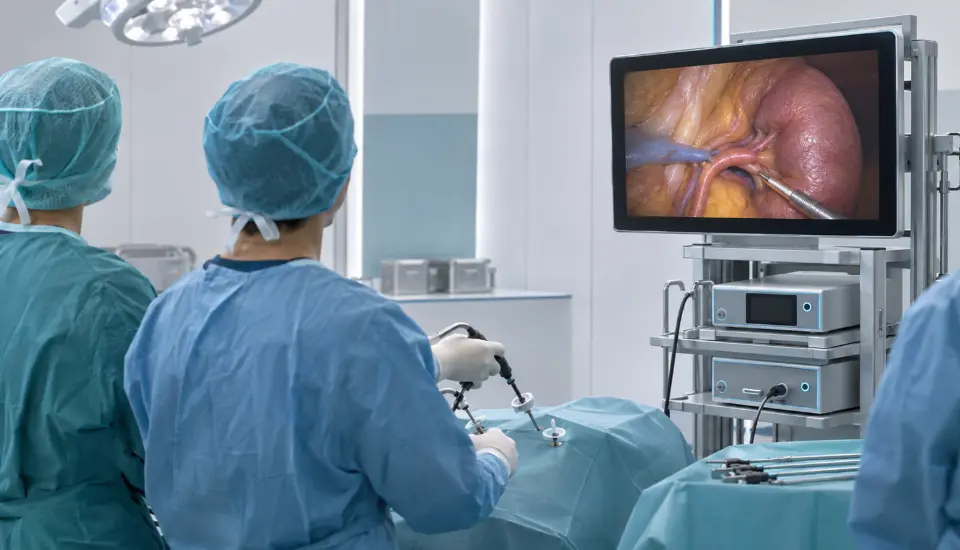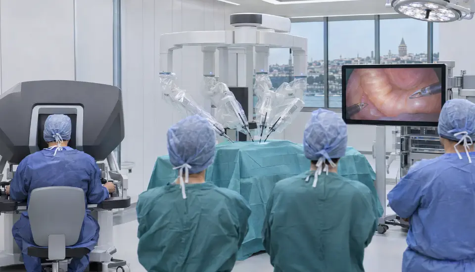
The hands-on day of the Robotic Colorectal Surgery Cadaver Course in Istanbul begins with a task that can determine how comfortably the rest of an operation proceeds: positioning the patient, placing the ports and docking the robot.
Each participant will perform these steps using the da Vinci Xi system before the group moves on to colorectal dissection. It is a practical starting point for a course that will eventually take surgeons from the right colon, across the splenic flexure and into the pelvis.
The course takes place on 5-6 December 2026 in Istanbul, Türkiye, combining an afternoon of theoretical teaching with a full day of cadaveric training. The surgical program includes robotic right hemicolectomy with complete mesocolic excision and central vascular ligation (CME/CVL), left colectomy, splenic flexure mobilization and total mesorectal excision (TME).
Participation is limited to just four surgeons.
This makes the program a private, small-group training experience rather than a conventional surgical workshop with a large number of delegates. Participants follow a planned rotation between the robotic console and bedside activities, working with a fresh-frozen cadaver under faculty supervision.
For surgeons interested in developing their robotic colorectal skills, the practical value lies in the details: finding the correct dissection plane, identifying vascular structures, managing the robotic arms and understanding how the approach changes as the operation moves from the abdomen into the pelvis.
Table of Contents
Robotic Colorectal Surgery Cadaver Course at a Glance
📌 Format: Theory, surgical video analysis and hands-on fresh-frozen cadaver lab
📅 Duration: 2 days, 5–6 December 2026
📍 Location: Istanbul, Türkiye
🤖 Robotic system: da Vinci Xi
🩺 Procedures: Robotic right hemicolectomy (CME/CVL), left colectomy, splenic flexure mobilization and TME
🧪 Training model: Fresh-frozen cadaveric dissection
👨⚕️ Group size: Private course limited to 4 participants
🎓 Certificate: Certificate of Participation, subject to course completion requirements
💳 Course fee: USD 4,500 per participant
💰 Registration deposit: USD 500
📞 Contact: For registration details and availability, contact our medical education team via WhatsApp at +90 537 977 89 84.
Why a Four-Participant Course?
One of the first questions surgeons ask about a robotic training program is how much time they will actually spend working with the system.
A long procedure list is attractive, but it does not necessarily tell participants what their experience in the laboratory will look like. When a group is large, much of the practical session can be spent observing other participants.
This December course has a maximum of four places.
The program includes a defined rotation between console and bedside responsibilities, allowing the group to work through the planned procedures in a closely supervised setting. It also gives the faculty an opportunity to discuss technique with a small number of participating surgeons.
The limited group size is particularly relevant to robotic training because several important tasks take place outside the console.
Docking, patient positioning, instrument exchange and bedside coordination all affect how smoothly the operation proceeds. A surgeon learning the robotic approach benefits from understanding these activities rather than concentrating entirely on the surgeon’s console.
During the cadaver lab, each participant is scheduled to perform positioning, port placement and docking for the right-colon setup. The subsequent exercises involve console and bedside rotation as the group progresses through the procedures.
The published program does not allocate a fixed number of console minutes to each physician. What it does establish is the small group size, the rotation arrangement and the surgical work planned for the day.
Learning the da Vinci Xi Before Starting the Dissection
The theoretical program begins on Saturday, 5 December, with registration and opening remarks.
The first teaching session is devoted to setting up the da Vinci Xi for colorectal surgery.
The faculty will review the main components of the system, including the patient cart, surgeon console and vision cart. Port placement is discussed for right-sided, left-sided and pelvic procedures, together with patient positioning, targeting and boom rotation.
These subjects may seem elementary to surgeons already familiar with robotic operating rooms. In practice, however, the arrangement of the robotic system becomes increasingly important when an operation involves several anatomical regions.
A right hemicolectomy and a pelvic TME do not place the same demands on port geometry or instrument movement. The surgeon has to consider how the robotic arms will approach the target and whether the working arrangement will remain suitable as the dissection progresses.
Instrument selection is also included, particularly the use of the needle driver and Firefly imaging.
The session addresses the bedside assistant’s responsibilities, ergonomics and emergency undocking procedures. These are essential parts of robotic operating room practice, even though they receive less attention than the dissection itself.
The first hour provides the technical foundation for the colorectal procedures discussed later in the afternoon.
Robotic Right Hemicolectomy: Vascular Anatomy and Dissection Planes
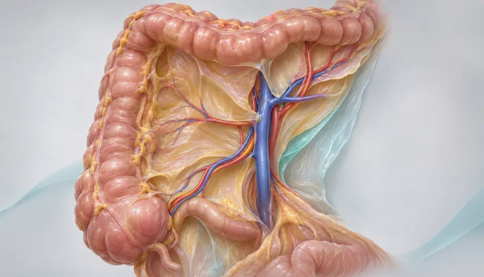
The next session, from 14:15 to 15:15, concentrates on robotic right hemicolectomy.
The teaching covers complete mesocolic excision and central vascular ligation, commonly referred to as CME/CVL.
A central part of this procedure is understanding the relationship between the mesocolon, the superior mesenteric vessels and the vascular branches supplying the right colon.
The program includes exposure of the superior mesenteric vein (SMV), identification of the ileocolic, right colic and middle colic vessels, and recognition of anatomical variations.
The gastrocolic trunk of Henle is another important structure discussed during the session.
Vascular anatomy is particularly relevant in right-sided colorectal surgery because the arrangement of these vessels is not identical in every patient. The surgeon must recognise the anatomy encountered during dissection rather than rely on a single expected branching pattern.
The course examines different operative approaches, including medial-to-lateral and cranial-to-caudal dissection.
Both approaches require an understanding of the appropriate embryological planes. During CME, the surgical objective is to work within the relevant mesocolic plane while respecting the surrounding anatomy.
Intracorporeal anastomosis techniques complete the right-colon session.
Recorded surgical videos are used throughout the teaching, with time for questions and discussion.
This prepares participants for Sunday’s practical work, when the same anatomical structures and dissection principles will be encountered in the cadaver lab.
Left Colectomy and the Challenge of the Splenic Flexure
After the afternoon break, teaching continues with left colectomy, splenic flexure mobilization and TME.
The session runs from 15:30 to 16:45.
For left-sided colorectal surgery, the vascular discussion moves to the inferior mesenteric artery (IMA) and inferior mesenteric vein (IMV).
The faculty will examine the relevant anatomy, vascular variations and approaches to nerve-preserving dissection.
Splenic flexure mobilization receives particular attention.
The program covers medial-to-lateral, lateral-to-medial and supramesocolic approaches. These techniques offer different ways to access and release the splenic flexure, depending on the operative circumstances.
Mobilization in this region requires careful orientation around the pancreas, mesocolon and adjacent structures. The selected approach may influence exposure and the sequence of dissection.
It is also a procedure where instrument positioning and traction can make the difference between a clear operative view and a difficult working angle.
During the theoretical session, the faculty will discuss these approaches using surgical recordings.
The following day’s cadaver exercises give participants an opportunity to work through the relevant anatomy while performing robotic dissection.
Total Mesorectal Excision and Autonomic Nerve Preservation
The theoretical program then moves into the pelvis.
Robotic total mesorectal excision is one of the three principal procedures covered during the course, alongside right hemicolectomy and left colectomy.
Pelvic dissection brings a different set of anatomical challenges.
The working space is narrower, the operative view changes as dissection progresses, and important autonomic nerve structures lie close to the intended surgical planes.
The December program includes pelvic exposure, mesorectal dissection planes and nerve preservation.
Particular attention is given to the hypogastric nerves, pelvic autonomic plexus and Denonvilliers’ fascia.
For surgeons performing TME, identifying and respecting the correct anatomical planes is central to the procedure. The relationship between the mesorectum, pelvic nerves and surrounding fascia must be understood throughout the dissection.
The faculty will also discuss anastomosis in a deep or narrow pelvis, including perfusion assessment using indocyanine green (ICG).
This subject forms part of Saturday’s theoretical teaching. The cadaver lab itself focuses on anatomical dissection and the scheduled robotic operative steps.
Video-based discussion gives participants an opportunity to examine the approach before working in the pelvic region during Sunday’s session.
Complications, Instrument Problems and Decisions at the Console
Saturday’s final teaching session begins at 17:00 and finishes at 18:15.
The focus is on technical difficulties and complications.
Bleeding control at the console is discussed alongside the prevention and management of ureteral, nerve and bowel injuries.
The faculty will also address instrument collisions and docking problems.
These problems are specific considerations in robotic surgery. The surgeon needs a clear understanding of the robotic arms and their working range, while the bedside team must be able to respond appropriately when adjustments are needed.
Conversion criteria form another part of the discussion.
The decision to change the operative approach requires judgement based on the circumstances of the procedure. The session provides an opportunity to discuss these decisions through surgical experience and case-based teaching.
An interactive question-and-answer session closes the theoretical day.
Sunday: A Full Day of Robotic Cadaveric Dissection
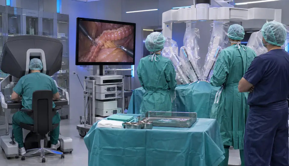
The second day begins at 08:00 with a laboratory briefing.
Participants are introduced to the characteristics of the fresh-frozen cadaver model, including the differences between cadaveric tissue and live surgery. Laboratory safety, instrument handling and the planned rotation between console and bedside activities are also covered.
Fresh-frozen cadaveric training offers an opportunity to examine surgical anatomy and practise dissection without operating on a patient.
There are limitations. A cadaver does not reproduce active bleeding, normal tissue perfusion or every response encountered during live surgery. The model is therefore most useful for anatomical orientation, instrument handling and the technical steps selected for the exercises.
The course uses this setting to work through the principal colorectal procedures in sequence.
Right-Colon Positioning and Docking
From 08:30 to 09:15, participants work on positioning, port placement and docking for right-colon surgery.
Each physician is scheduled to perform the setup.
The session includes camera handling and effective use of the third robotic arm, with attention to how the instruments are positioned for the dissection ahead.
This gives participants practical exposure to the setup principles introduced on Saturday.
Robotic Right Hemicolectomy: 09:15–12:00
The morning’s main dissection session is devoted to robotic right hemicolectomy with CME/CVL.
Participants work on identifying the anatomy, selecting instruments and performing medial-to-lateral dissection.
The exercises include SMV exposure and dissection involving the ileocolic, right colic and middle colic vessels.
The gastrocolic trunk of Henle and hepatic flexure mobilization are also covered.
Needle-driver use and intracorporeal anastomosis are included in the morning program.
The faculty supervise the planned exercises as participants rotate between console and bedside roles.
Lunch follows from 12:00 to 12:45.
Re-Porting and Left Colectomy
The afternoon begins with a 30-minute session devoted to re-porting and re-docking.
The program addresses the setup for splenic flexure and pelvic work, including the planned docking configuration.
At 13:15, the group moves into left colectomy and splenic flexure mobilization.
The practical exercises include nerve-preserving IMA dissection and ligation, IMV ligation and identification of the relevant vascular anatomy.
Participants also work on medial-to-lateral dissection and the different approaches to splenic flexure mobilization.
The session continues until 15:30.
Robotic Total Mesorectal Excision: 15:45–17:45
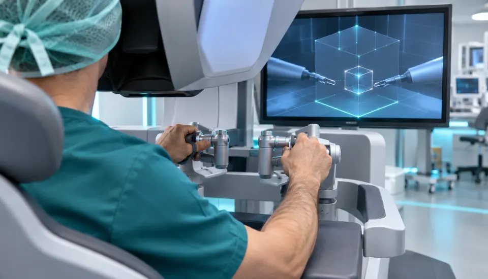
The final cadaveric session is devoted to TME.
The scheduled exercises cover posterior, lateral and anterior pelvic dissection.
Participants identify the hypogastric nerves and pelvic plexus, examine Denonvilliers’ fascia and continue the dissection towards the pelvic floor.
Distal rectal transection is also part of the program.
This is the most pelvis-focused portion of the course and brings together several subjects discussed during Saturday’s theoretical teaching.
Working through the dissection in a cadaver model allows the group to examine the anatomical planes and the spatial relationships between the rectum, mesorectum and surrounding structures.
The laboratory session ends at 17:45.
Participant feedback and the certificate ceremony follow, with the course concluding at 18:15.
Who Is This Robotic Colorectal Surgery Course For?
The course is intended for surgeons interested in advanced robotic colorectal procedures.
It may be particularly relevant to practising general or colorectal surgeons who want additional exposure to da Vinci Xi setup, colorectal vascular anatomy, robotic dissection and pelvic surgery.
Surgeons developing their robotic practice may find the positioning, docking and instrument-handling components useful. Physicians who already have robotic experience may be more interested in the CME/CVL dissection, splenic flexure approaches or anatomical details of TME.
The course also covers intracorporeal anastomosis and needle-driver techniques, which may be of interest to surgeons seeking further practice with robotic suturing.
Given the advanced procedural content, prospective participants should confirm that the course matches their current surgical experience. The supplied program does not specify formal prerequisites.
With only four available places, the course is designed as a small-group practical training experience. It does not, however, replace supervised clinical training or establish independent competence in the listed procedures.
Course Fee and Registration
The participation fee for the Robotic Colorectal Surgery Cadaver Course is USD 4,500 per physician.
The fee includes access to the two-day scientific and practical program, scheduled hands-on training with the da Vinci Xi Surgical System, and course-related educational materials and resources.
Membership with access to eligible member benefits and future course opportunities is also included.
A Certificate of Participation is issued subject to completion of the course requirements.
How to Register
For registration details, availability and the official registration form, please contact our medical education team via WhatsApp at +90 537 977 89 84.
Four Places for the December 2026 Cadaver Course
The Robotic Colorectal Surgery Cadaver Course in Istanbul on 5–6 December 2026 brings together several demanding colorectal procedures within a private training format limited to four participants.
The program begins with da Vinci Xi setup and operative video discussions, followed by a cadaver lab covering right hemicolectomy, left colectomy, splenic flexure mobilization and TME.
The small group and planned console-and-bedside rotation are important parts of the course structure. So is the progression from right-sided vascular dissection to left-colon surgery and the anatomical challenges of the pelvis.
Surgeons considering participation can review the detailed program and request registration information from the course organizers.
Further articles about specialist surgical training and upcoming medical education programs in Istanbul are available in the MedClinics Medical News & Blog section.
Frequently Asked Questions
When and where is the Robotic Colorectal Surgery Cadaver Course taking place?
The course will be held in Istanbul, Türkiye, on 5-6 December 2026. Saturday is dedicated to theoretical teaching and surgical video discussions, while Sunday focuses on the cadaver lab.
Is this a private course?
Yes. Participation is limited to four physicians, making it a private, small-group surgical training program.
Which robotic system will participants use?
The course includes scheduled hands-on training with the da Vinci Xi Surgical System, covering positioning, docking and robotic colorectal techniques.
Which operations are included in the cadaver lab?
The program covers robotic right hemicolectomy with CME/CVL, left colectomy, splenic flexure mobilization and total mesorectal excision.
What is the participation fee?
The course fee is USD 4,500 per participant. A USD 500 deposit is required to secure registration, with the remaining balance payable according to the agreed arrangements.
Is accommodation included in the course fee?
No. Accommodation is not included in the standard participation fee. An optional two-night accommodation and transfer package is available for USD 350 per participant.
Will participants receive a certificate?
Yes. Participants receive a Certificate of Participation, subject to meeting the course completion requirements.