MedClinics News & Blog
SkinTE treatment was tested in people with diabetic foot ulcers that had already remained open for an average of nine to ten months.
The Phase 3 study included 120 patients. Half received SkinTE alongside standard wound treatment; the other half continued with standard treatment alone. At week 12, complete wound closure had been achieved in 32 patients in the SkinTE group and 16 in the comparison group.
SkinTE is made from the patient’s own skin. A clinician takes a small biopsy from a healthy area, and the tissue is processed into a multicellular preparation for the ulcer. No donor cells are involved.
SkinTE is an autologous skin therapy within regenerative medicine, although it is not a conventional stem cell treatment. The material placed on the ulcer contains several types of skin cell as well as extracellular matrix.
Robert S. Kirsner, MD, PhD, of the University of Miami, presented the COVER DFUS II results at the Symposium on Advanced Wound Care Fall meeting in October 2026. PolarityBio sponsored the trial and is developing SkinTE. For now, the most detailed information comes from the conference presentation and the registered study record. The journal article is still to come.
Table of Contents
The Trial Selected Hard-to-Heal Diabetic Foot Ulcers
There is rarely one reason why a diabetic foot wound stays open.
A patient may not feel that a shoe is rubbing or that one area of the foot is carrying too much pressure. The same spot is then disturbed with every step. Blood supply may be limited, and an open wound gives bacteria an opportunity to enter. These problems often overlap rather than arriving one at a time.
Before patients entered the randomized stage of COVER DFUS II, they received two weeks of standard wound care. Anyone whose ulcer became more than 30% smaller during that period was excluded.
This left the researchers with wounds that were showing little progress.
All were Wagner grade 1 ulcers measuring between 1 and 15 square centimetres. They had passed through the skin but had not reached the deeper structures involved in the higher Wagner grades.
The patients’ histories reveal how serious their foot disease had become. Around 60% had undergone an earlier amputation. Some had previously experienced infection or osteomyelitis.
The Patient Supplied the Starting Material
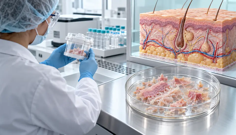
The SkinTE treatment begins with a small biopsy taken from the patient’s healthy skin. The biopsy went through the surface layer and into the dermis, giving the manufacturer more than a thin sample of epidermis to work with.
Processing did not reduce that tissue to one chosen cell. The material returned to the clinic as many small units containing elements of the original skin.
Among them were keratinocytes, fibroblasts and cells associated with blood vessels and hair follicles. Extracellular matrix remained in the preparation too.
In normal skin, those parts are closely connected. The surface depends on keratinocytes, while fibroblasts and matrix help give the dermis its structure. Small vessels, follicular tissue and the signals travelling between cells add to the local environment.
SkinTE brings some of that mixture to the ulcer. What follows inside the wound is still partly a black box. The trial tells us whether the skin surface eventually closed; it does not reveal which cells lasted, which disappeared or which had the greatest influence on repair.
Complete Wound Closure Was the Main Endpoint
COVER DFUS II was conducted at 17 centres in the United States. The 120 participants were assigned at random to two equal groups.
Sixty received SkinTE plus standard wound care. The other 60 continued with standard care alone.
Both groups still needed the basics of diabetic foot care: regular review, wound protection, debridement where necessary and relief of pressure from the affected area.
The main endpoint was complete closure at week 12. A reduction in wound size was not enough. The surface had to be covered with new epithelium, without drainage and without the continuing need for a dressing.
That choice kept the result close to the patient’s experience. A smaller wound may be encouraging, but it is still open. A closed wound changes dressing needs and reduces the exposed surface through which infection can enter.
Most patients received SkinTE only once. A second application was allowed, but it was used in 18.3% of the treatment group.
Complete Wound Closure Was Twice as Common
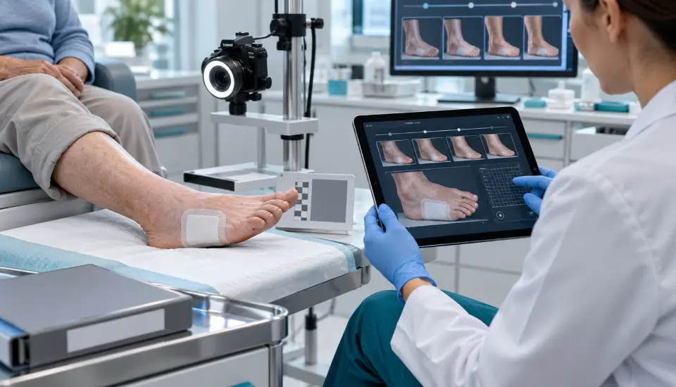
At week 12, complete wound closure had been achieved in 32 of the 60 patients receiving SkinTE. Standard care produced 16 closed wounds among the other 60 patients.
In percentage terms, the two results were 53.3% and 26.7%. The statistical analysis gave a p-value of 0.0014, meeting the trial’s primary endpoint.
Median time to closure in the SkinTE group was 85 days. A median could not be calculated for standard care because fewer than half of those ulcers closed.
The result can be understood without relying on a specialist scoring system. Twice as many wounds were closed at the end of 12 weeks when SkinTE had been used.
Still, 28 ulcers in the treatment arm had not closed. Perhaps some were larger, older or affected by poorer circulation. The presentation does not give enough patient-level detail to know. That question will have to wait for the longer paper.
Fewer Wound Complications Were Reported
The results for infection moved in the same direction as wound closure.
Infection of the study ulcer was reported in 8.5% of the SkinTE group and 25% of the control group. Osteomyelitis occurred in 1.7% and 15%, respectively.
Three patients receiving standard care underwent an ulcer-related amputation. None was reported among patients treated with SkinTE.
Earlier closure could plausibly reduce the time during which bacteria can enter the wound. The complication findings came from a limited number of cases, however. In groups of 60, each patient shifts the result by roughly 1.7 percentage points.
The meeting report gives the totals, not the story behind every event. When did the infections occur? Had those wounds been open longer? What led to each amputation? The full study report will be more useful for answering those questions than the percentages alone.
No adverse event was directly attributed to SkinTE in the conference presentation. It would also be helpful to see how the biopsy sites healed, since every patient receiving SkinTE had a second, deliberately created wound.
Why SkinTE Is an Autologous Skin Therapy
The biopsy and the person receiving it are the same, which makes SkinTE autologous. Here, “autologous” describes where the tissue came from; it does not define a particular kind of cell.
An MSC treatment usually starts by selecting mesenchymal stromal cells and growing them under controlled laboratory conditions. SkinTE takes another route. It keeps different parts of healthy skin together instead of building the product around a single population.
There may be progenitor cells within the preparation, especially around the follicular tissue. They are not separated out, multiplied and given back as a stand-alone stem cell dose.
Nor did the trial add an exosome preparation. Cells in SkinTE may release extracellular vesicles after the product reaches the wound, just as living cells do elsewhere in the body. Those naturally released signals are not the same as manufacturing and administering purified exosomes.
As an autologous skin therapy, SkinTE uses tissue obtained from the patient rather than donor cells. Its place within regenerative medicine comes from the use of viable skin tissue to encourage a chronic wound to resume repair.
SkinTE Treatment Was Added to Standard Wound Care
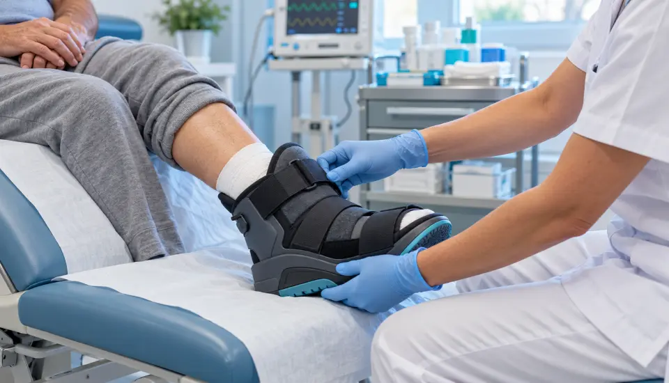
The SkinTE treatment was studied alongside standard care for diabetic foot ulcers, including pressure relief, wound review and debridement when required.
The particular offloading method would depend on the patient and the site of the ulcer. A cast may suit one case, while another patient needs a removable boot or altered footwear.
Circulation and infection require their own decisions. If the foot is receiving too little blood, vascular care may be necessary. An infection cannot be left for the regenerative product to solve.
COVER DFUS II tested SkinTE within that wider package. Repeated pressure, untreated infection or severe ischemia would change the conditions considerably.
The wounds were also limited to Wagner grade 1. Deeper ulcers involving tendon, joint or bone sit outside the evidence reported here.
What the Conference Results Leave Unanswered
The headline result is easy to understand, but the meeting presentation cannot carry the detail of a journal paper.
One important question is how closure was confirmed. Wound photographs and measurements involve some judgement, particularly near the point of complete healing. Knowing who reviewed them—and whether that person knew which treatment had been given—will help readers assess the result.
Patients were followed for 24 weeks, twice as long as the main endpoint period. Those later visits may reveal whether closure lasted. With neuropathy and abnormal pressure still present, an apparently healed ulcer can open again.
The published analysis should also show what happened when visits were missed or a patient left the study. It may identify patterns among patients whose wounds closed and those whose wounds did not.
PolarityBio sponsored COVER DFUS II and manufactures SkinTE. That is a common arrangement for a product approaching regulatory review. Once the complete methods and data are public, wound-care specialists outside the company can examine the work in more depth.
FDA Review Has Begun
PolarityBio has asked the FDA for permission to market SkinTE through a Biologics License Application.
The company announced that the application had entered Priority Review. The FDA’s current target for completing the review is 16 March 2027.
For now, this autologous skin therapy remains investigational while the FDA reviews the application. Priority Review moves the application through a shorter timetable, but the final decision still depends on the agency’s assessment of clinical evidence, manufacturing and safety.
Routine use would involve more than applying the product at a clinic visit. Healthy skin must first be collected and sent away. The processed material then has to return to the wound centre at the right time. Price, insurance coverage and access to clinics familiar with the process will shape how practical the treatment becomes.
The trial’s two-week run-in period may also offer a clue about patient selection. It allowed ordinary wound care a chance to work before SkinTE was used. In practice, a similar approach could help identify ulcers that have genuinely become stuck.
What Can Be Said Today
The central finding from COVER DFUS II is encouraging: 32 of 60 SkinTE-treated ulcers closed within 12 weeks, compared with 16 of 60 ulcers receiving standard care alone.
Lower rates of infection, osteomyelitis and amputation were reported as well, although these results involved fewer events.
The SkinTE treatment has therefore produced a positive randomized Phase 3 result in a difficult group of chronic diabetic foot ulcers. The upcoming journal paper should show how durable and consistent that result was.
For now, this autologous skin therapy is a promising option that could eventually sit alongside established wound care. The next milestones are publication of the complete study, longer follow-up and the FDA’s decision.
Frequently Asked Questions
How was the SkinTE treatment used?
The SkinTE treatment was added to standard diabetic wound care and compared with standard care alone.
How many patients took part?
COVER DFUS II included 120 patients treated at 17 centres in the United States.
How often was complete wound closure achieved?
Complete wound closure occurred in 32 of 60 SkinTE-treated ulcers and 16 of 60 ulcers in the comparison group.
Is SkinTE an autologous skin therapy?
Yes. SkinTE is an autologous skin therapy because each preparation is produced from the treated patient’s own healthy skin
Is SkinTE a stem cell treatment?
No single stem cell population was isolated or expanded. The product retained several kinds of skin cell together with extracellular matrix.
Were exosomes administered?
No separate exosome product was used in the trial.
Is SkinTE already approved?
No. The FDA is reviewing the company’s application, and SkinTE remains investigational.
Has the Phase 3 study been peer reviewed?
The results have been presented at a scientific meeting. The complete journal article has not yet appeared.
Sources
- HMP Global Learning Network – Phase 3 Trial Demonstrates Autologous Cutaneous Multicellular Units Improve Wound Closure Rates
- ClinicalTrials.gov – COVER DFUS II, NCT06140303
- PolarityBio – COVER DFUS II Phase 3 Results Presented at SAWC Fall 2026
- International Working Group on the Diabetic Foot – 2023 Wound Healing Guideline
- IWGDF/IDSA – Guidelines on the Diagnosis and Treatment of Diabetes-Related Foot Infections
This article is for informational purposes and does not replace professional medical advice, diagnosis or treatment.