MedClinics News & Blog
Donor kidney preservation usually means cooling the organ and limiting damage before transplantation. In this study, researchers tried something different: they added MSC-derived extracellular vesicles directly to the liquid circulating through donated human kidneys. The experimental treatment was not given to a patient or administered through an intravenous infusion.
All eight kidneys had already been declined for transplantation. Four underwent standard machine preservation, while the remaining four received the same procedure with extracellular vesicles added. After four hours, the EV-treated kidneys showed signs that their cells had handled the preservation period better.
It is an early finding, but an intriguing one. Instead of using regenerative medicine after an organ has been transplanted, researchers are asking whether a vulnerable donor organ could be supported before it reaches the patient.
Table of Contents
Donor Kidney Preservation Outside the Body
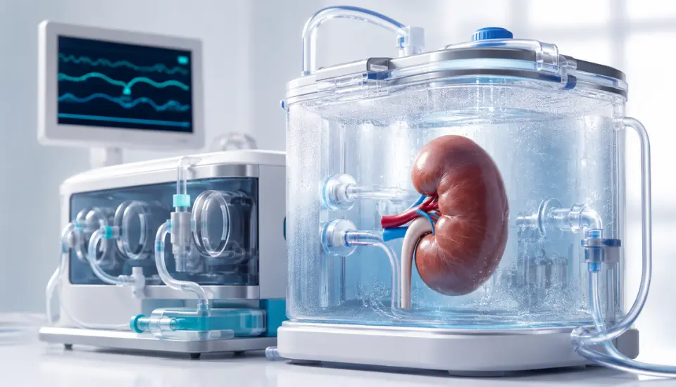
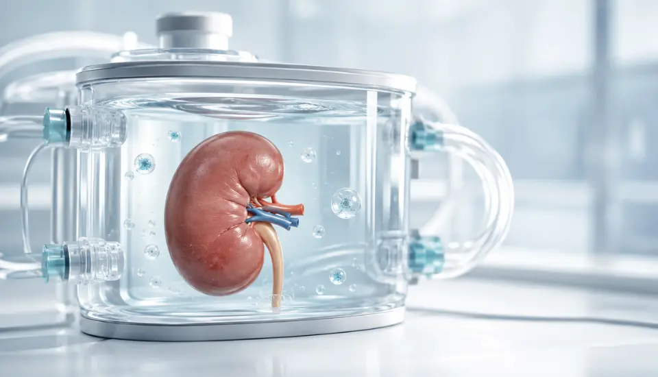
Donor kidneys are cooled after retrieval to slow their metabolism and limit tissue damage. Some are also connected to a perfusion machine, which circulates preservation fluid through the organ.
The kidneys in this study underwent hypothermic oxygenated perfusion, usually shortened to HOPE. The circulating solution was kept cold and supplied with oxygen while the kidneys remained outside the body.
This creates more than a way to store an organ. It also offers a brief period during which researchers can observe the kidney and test whether a biological intervention changes its condition.
The study involved kidneys that were considered unsuitable for clinical transplantation because of marginal donor characteristics and substantial tissue damage. They were divided evenly between two groups. Four received HOPE alone; four received HOPE with MSC-derived extracellular vesicles in the perfusion fluid.
Using hypothermic oxygenated perfusion in both groups allowed the researchers to compare the effect of the vesicles without changing the underlying preservation method.
Tissue was collected before and after the four-hour procedure. The researchers then examined the proteins, genes and structural markers present in each kidney.
No transplant recipients took part, and none of the kidneys were implanted afterwards.
Why Use Extracellular Vesicles Instead of MSCs?
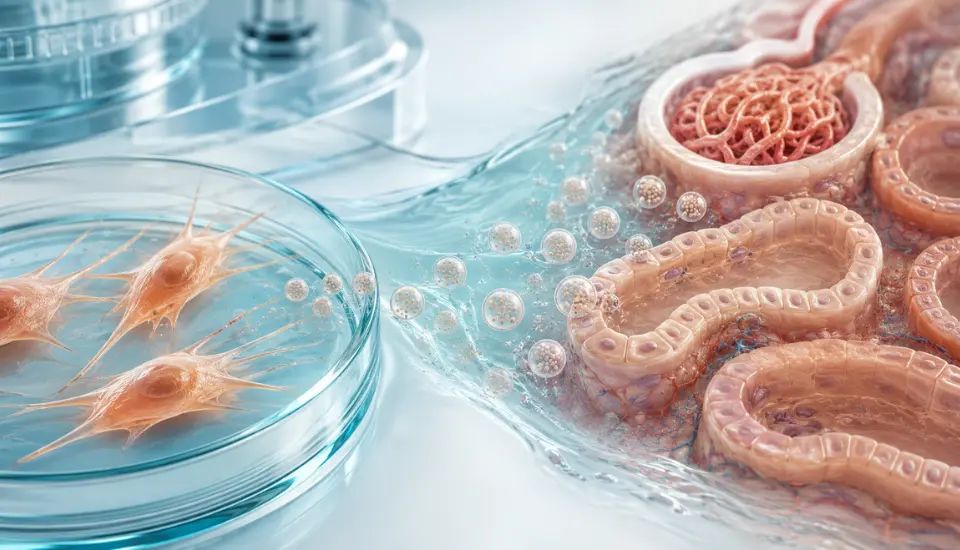
Mesenchymal stromal cells, widely known as MSCs, release small membrane-covered particles into their surroundings. These extracellular vesicles carry proteins, lipids and other biological material from one cell to another.
They are part of the way cells communicate. Depending on their contents, they may affect inflammation, cellular stress and tissue repair.
For years, researchers assumed that many benefits associated with MSC therapy came from the cells settling in damaged tissue and developing into replacement cells. The picture now appears more complicated. Some effects may be driven by the messages MSCs release, including those transported inside extracellular vesicles.
The kidney study explored this cell-free approach. No living stem cells were placed in the organs. The researchers used material released by MSCs and introduced it during machine perfusion.
The paper refers to the preparation as MSC-derived extracellular vesicles. Calling it an “exosome treatment” would be less precise because extracellular vesicles include several types of particles and the product was not described simply as purified exosomes.
What Was Different After Four Hours?
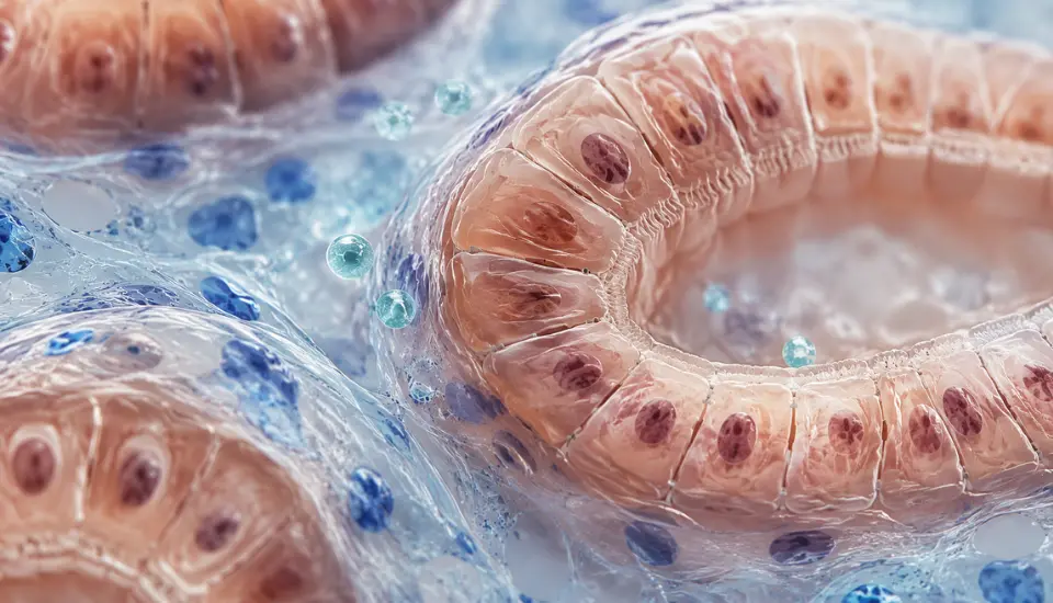
The strongest results came from the biological condition of the kidney tissue.
One of the markers studied was p16. It is commonly associated with cellular senescence – a state in which cells stop dividing normally and may begin contributing to inflammation and tissue dysfunction. Levels of p16 were lower in kidneys perfused with extracellular vesicles.
The opposite pattern appeared with Ki-67, a marker linked to cell proliferation. Ki-67 increased in the EV-treated tissue.
Researchers also examined E-cadherin, a protein that helps neighbouring cells remain connected and supports the structure of the kidney tubules. Its preservation was better in the kidneys that received extracellular vesicles.
A wider analysis of the tissue proteins found changes in pathways involved in antioxidant defence, cell survival, metabolism and structural stability. Some proteins connected with cellular ageing became less active, while survival-related signals increased.
These are useful findings, although they should not be mistaken for proof that the kidneys were restored. The study shows that the cells responded differently during preservation. It does not show that the organs recovered normal kidney function.
Why the Use of Human Kidneys Is Significant
A cell culture can reveal how one cell type reacts to an experimental treatment. An animal model can show how that treatment behaves inside a living body. Neither fully reproduces the structure of a human kidney.
Here, the researchers worked with the complete organ: its blood vessels, tubules, connective tissue and different cell populations were all present. That gives the findings more clinical relevance than a laboratory experiment using isolated cells alone.
The method is also practical for donor kidney preservation. Machine perfusion is already designed to circulate fluid through a donor kidney. Adding a biological product to that fluid would allow the organ to be treated directly, without first administering the product to the transplant recipient.
This does not mean the approach is ready to use. It does mean that, if later studies are successful, the treatment could fit into an existing stage of the transplantation process.
The Missing Part: Transplantation
There is one major question this study could not answer: would the treated kidneys have worked after transplantation?
The organs were not connected to a human blood supply after perfusion. Researchers did not observe urine production or long-term filtration, and there was no opportunity to measure delayed graft function, rejection or survival in a recipient.
The sample was also very small. With four kidneys in each group, unusual characteristics in a single organ could have a noticeable effect on the overall result.
There is another challenge on the EV side. The biological contents of extracellular vesicles depend on where the MSCs came from and how they were grown. Isolation, storage and dosing methods can change the final preparation as well. A repeatable manufacturing process will be needed before different studies can be compared fairly.
For those reasons, this paper should not be presented as evidence that MSC-derived extracellular vesicles can already rescue kidneys that have been rejected for transplant.
What it provides is a starting point: damaged human kidney tissue appeared to enter a more protective biological state when exposed to the vesicles during machine perfusion.
A Different Place for Regenerative Medicine
Regenerative medicine is often described as something given directly to a patient. This research takes a different route. The target is the donated organ during the hours between retrieval and possible transplantation.
That is a particularly interesting window. The kidney is accessible, its circulation can be controlled, and researchers can deliver a product without exposing the recipient’s entire body to it at the same time.
Future experiments will need more kidneys, longer observation and stronger measures of organ function. Eventually, the question must be tested in a carefully controlled transplant setting.
Until then, the result remains promising biology rather than a new transplantation method. Still, it gives researchers a credible reason to continue exploring MSC-derived extracellular vesicles as part of donor kidney preservation.
Frequently Asked Questions
Was this a study in kidney transplant patients?
No. Eight donated kidneys were studied outside the body. No patients received the extracellular vesicles.
Were living stem cells used?
No. The researchers used extracellular vesicles released by MSCs. Living MSCs were not introduced into the kidneys.
Did the treated kidneys become suitable for transplantation?
The study did not assess that. The kidneys showed favourable biological changes, but they were not transplanted and their long-term function was not tested.
What did the extracellular vesicles appear to do?
They were associated with lower levels of a cellular-senescence marker, greater expression of a proliferation marker and better preservation of a protein important to kidney-tubule structure.
Is this method available clinically?
No. Using MSC-derived extracellular vesicles during donor kidney perfusion remains experimental.
Sources
- Proteomic Profiling of Discarded Human Marginal Kidneys Undergoing Hypothermic Oxygenated Perfusion: A Comparison of Treatment With Versus Without MSC-Derived Extracellular Vesicles
- ClinicalTrials.gov – NCT03837197: Strategies to Improve Function of Marginal Grafts and Recover Discarded Grafts in Liver and Kidney Transplantation
This article is for informational purposes and does not replace professional medical advice, diagnosis or treatment.