MedClinics News & Blog
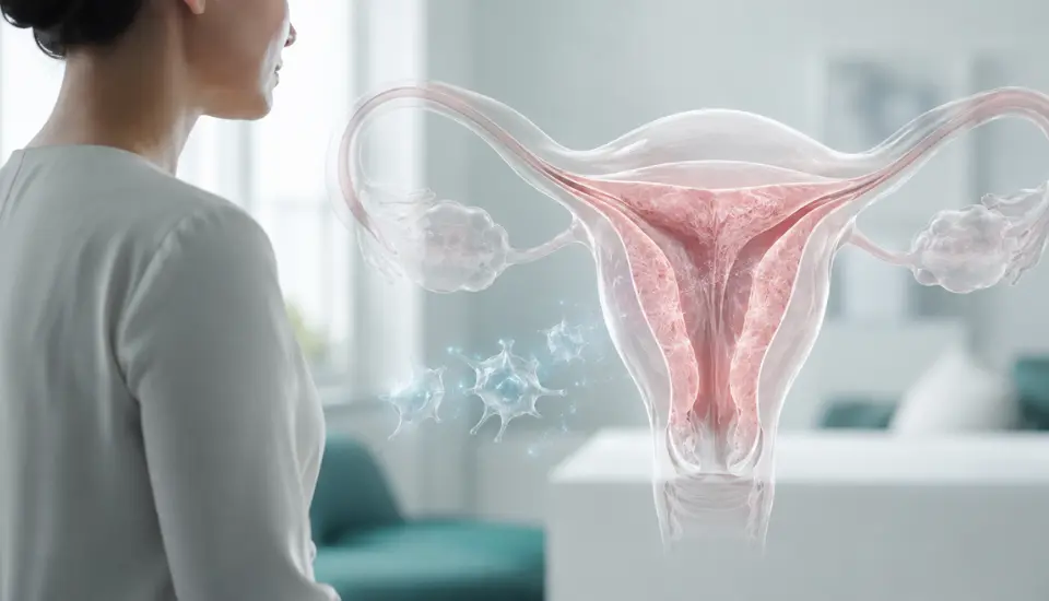
Stem cell therapy for intrauterine adhesions, including severe cases associated with Asherman syndrome, is often discussed as a future possibility. A paper published on August 7, 2026, gives the field something more concrete: 60 women, a control group, blinded ultrasound measurements and follow-up through embryo transfer.
The women had severe scarring inside the uterus. They also had good-quality frozen embryos, but their transfers had been cancelled because the endometrium remained too thin. Following treatment, the uterine lining grew more in women who received bone marrow-derived mesenchymal stem cells. Clinical pregnancies were also recorded more frequently in this group.
The live birth figures require more caution. Nine women in the stem cell group gave birth, compared with five in the control group. The study was not large enough to confirm that the difference resulted from the treatment.
Even with that uncertainty, the paper adds useful human evidence to an area that has long depended on small, uncontrolled studies.
Table of Contents
When Removing Scar Tissue Is Not Enough
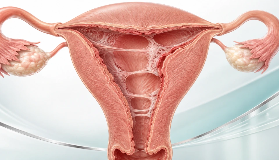
Intrauterine adhesions are bands of scar tissue that form within the uterus. The term Asherman syndrome is commonly used when they cause symptoms such as light or absent periods, infertility, repeated miscarriage or pelvic pain.
The endometrium—the tissue lining the uterine cavity—normally rebuilds itself every month. Hormones guide its growth and prepare it for embryo implantation. Much of the surface is shed during menstruation, then restored from the deeper basal layer.
An injury that reaches this deeper layer can interrupt that renewal. Healthy tissue may gradually be replaced by fibrosis. In some women, scar tissue connects opposite sides of the uterus and reduces the usable space inside the cavity.
These adhesions frequently develop after procedures related to miscarriage, pregnancy or delivery. Uterine surgery and infection can also play a role.
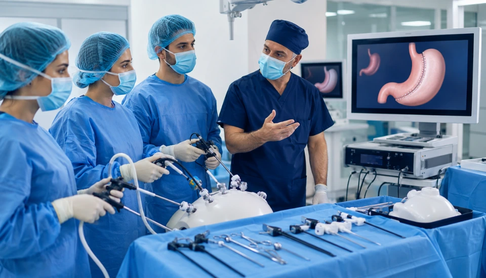
Treatment usually begins with hysteroscopy. Using a small camera, the surgeon locates and divides the adhesions. Hormone therapy is then prescribed to encourage the endometrium to grow over the treated area.
The difficult cases are those in which the cavity has been reopened, yet the lining still responds poorly. Further surgery cannot create healthy endometrial tissue by itself. Adhesions may return, and repeated procedures can add to an already complicated situation.
For women undergoing IVF, this can lead to a frustrating pause. Embryos are ready, while the uterine lining never reaches the point at which the medical team feels comfortable proceeding with transfer.
The Women Enrolled Had Severe Endometrial Damage
The new clinical study was conducted at the Sixth Affiliated Hospital of Sun Yat-sen University in China between July 2020 and December 2024.
Researchers enrolled 60 women aged 20 to 38. Severe intrauterine adhesions had been confirmed by hysteroscopy, and each participant had an endometrial thickness below 7 millimeters. All had undergone IVF or ICSI and had at least two good-quality frozen blastocysts.
The participants were assigned to two groups of 30. Everyone underwent hysteroscopic treatment, followed by two cycles of estrogen and progesterone. Low-dose aspirin was also included in the care protocol.
Women in the investigational group were additionally assigned to receive bone marrow-derived mesenchymal stem cells. Two withdrew before treatment, leaving 28 who underwent the cell procedure.
The stem cells came from the bone marrow of a healthy donor. They were expanded and checked under controlled manufacturing conditions. Each participant received one million cells per kilogram of body weight.
A catheter was introduced through the femoral artery and guided towards the arteries supplying the uterus. The cell suspension was then released through both uterine arteries.
The choice of delivery route is an interesting part of the study. A uterus affected by extensive adhesions may not distribute material placed directly inside the cavity evenly. By approaching through the blood vessels, the research team hoped to reach the deeper tissue and microvascular network involved in endometrial growth.
Because there was no sham catheter procedure, patients and treating doctors knew which treatment had been assigned. The clinicians responsible for ultrasound measurements remained unaware of the assignments. Embryologists involved in the later transfers were also blinded.
The Difference Was Less Than One Millimeter
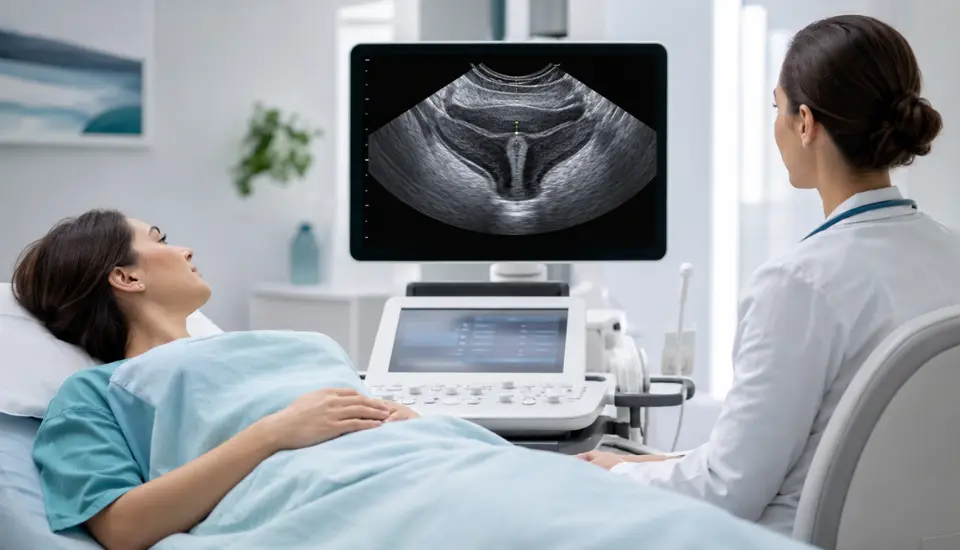
After treatment, average endometrial thickness reached 6.81 millimeters in the stem cell group and 6.42 millimeters in the control group.
The researchers also calculated how much the lining had grown from its starting measurement. The average increase was 0.79 millimeters following stem cell treatment and 0.20 millimeters with standard care alone.
At first glance, both numbers are small. Their clinical setting gives them more weight. These women had already experienced embryo-transfer cancellations because their endometrium did not grow sufficiently. A relatively modest increase could help an individual patient pass the threshold used to decide whether transfer can proceed.
Still, endometrial thickness is only one measurement. Ultrasound cannot fully describe blood flow, inflammation, fibrosis or the tissue’s biological readiness for implantation. Women can become pregnant with a thin endometrium, while an apparently adequate measurement does not guarantee success.
Here, the result shows that the lining responded more strongly in the group receiving stem cell therapy for intrauterine adhesions. Pregnancy follow-up provided the next piece of information.
More Clinical Pregnancies Followed Treatment
Embryo transfer was allowed once the endometrium reached at least 6 millimeters and the other study requirements had been met. Transfers were cancelled when the lining remained below this level, severe adhesions returned or fluid was seen inside the uterus.
Among women who proceeded with embryo transfer, 13 in the stem cell group developed a clinical pregnancy. Seven women became pregnant in the control group. The reported rates were 50% and 23.3%.
A clinical pregnancy required a gestational sac to be visible on ultrasound. The difference between the groups reached statistical significance.
The birth figures moved in the same direction. Nine women in the stem cell group and five in the control group had a live birth, corresponding to rates of 34.6% and 16.7%. In statistical terms, this result remained inconclusive. The wide range of possible outcomes means that chance cannot be excluded.
Miscarriage occurred in four of the 13 pregnancies following stem cell treatment and in two of the seven control pregnancies. With only 20 pregnancies altogether, the study offers little information about whether treatment had any effect on miscarriage risk.
The pregnancy rate is worth following in future research. Live birth needs to become the main measure in a larger study before the procedure can be judged as a fertility treatment.
The Tissue Findings Pointed Towards Blood-Vessel Repair
The researchers also examined tissue samples and performed laboratory experiments to understand the clinical findings.
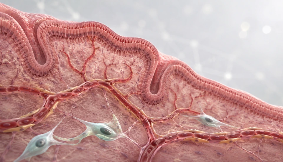
Endometrial samples associated with stem cell treatment had a greater density of small blood vessels. Activity also increased in biological pathways linked to angiogenesis, including VEGF and NOTCH signaling.
This is relevant because the endometrium depends on an active blood supply. Its vessels change throughout the menstrual cycle as the lining grows and prepares for implantation. Severe scarring can leave this network sparse or disorganized.
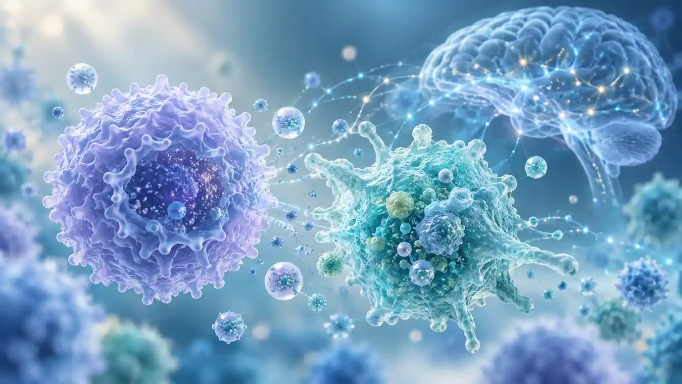
Another part of the analysis focused on pericytes. These cells sit around small blood vessels and help stabilize them. In laboratory conditions influenced by mesenchymal stem cells, pericytes showed greater movement and closer interaction with endothelial cells.
The results suggest that the donor cells may have altered the local repair environment through the substances they released. Improved vascular support could then have helped the patient’s own endometrial tissue respond to hormones.
There are gaps in this explanation. The researchers did not follow the infused cells inside each participant, and the gene analysis relied on a small set of tissue samples. Some pre- and post-treatment samples came from different women. The vascular findings should be viewed as clues about the mechanism.
Safety Was Reassuring During the Available Follow-Up
No serious adverse event was attributed to the bone marrow-derived mesenchymal stem cells or their infusion.
One participant developed bruising and a small hematoma where the catheter entered the femoral artery. It disappeared within one week and did not require further treatment.
The researchers reported no fevers, infections, allergic reactions or acute infusion problems. Blood counts, coagulation profiles, kidney tests and liver tests remained within normal ranges.
No ectopic pregnancies occurred. The investigators also recorded no major maternal complication linked to the procedure and no congenital abnormalities among the babies with available follow-up.
These observations cover only 28 treated women. A rare reaction might not appear until the procedure has been studied in a much larger population. Follow-up of future pregnancies and children will be especially important for any regenerative treatment used in reproductive medicine.
Where the Evidence Stands
The study has several features that improve confidence in its main result. Participants were assigned to treatment groups before the intervention. Both groups received the same hysteroscopic and hormonal care, and ultrasound measurements were assessed by clinicians who did not know which treatment had been given.
The patient group was narrowly defined. Participants were younger than 39, had adequate ovarian reserve and already had frozen embryos of suitable quality. Many people with intrauterine adhesions will not share that exact profile.
The open-label design also leaves some room for bias. Two women withdrew before receiving the stem cell infusion, and two others did not complete the post-treatment ultrasound assessment. The researchers used a conservative method to account for missing measurements in the primary analysis.
A further consideration is the delivery procedure. Arterial catheterization is more demanding than a local infusion into the uterine cavity. Its possible advantage must eventually be weighed against the expertise, monitoring and resources it requires.
For now, the most dependable finding is the increase in endometrial thickness. The pregnancy result adds clinical relevance and supports continued research. The live birth data remain preliminary.
What Researchers Need to Examine Next
A multicenter study would show whether these results can be reproduced by different medical teams. It should enrol enough women to use live birth as the primary outcome and should continue monitoring mothers and children after delivery.
Durability is another open question. The paper does not yet tell us whether the increase in endometrial growth persists across later cycles or whether it affects the recurrence of adhesions.
Researchers may also compare uterine artery infusion with less invasive delivery methods. If similar results can be achieved without arterial catheterization, that would influence how practical the approach becomes.
Stem cell therapy for intrauterine adhesions is currently an experimental option. It requires donor cells, specialized manufacturing and an interventional procedure. The new study does not establish it as routine fertility care.
It does bring the field closer to answering a clinically useful question for women with Asherman syndrome: can stem cell therapy for intrauterine adhesions help rebuild an endometrium that has remained thin after surgery and hormone treatment?
Frequently Asked Questions
What are intrauterine adhesions?
They are areas of scar tissue inside the uterus. Extensive adhesions may damage the endometrium, reduce the uterine cavity and interfere with menstruation, embryo implantation or pregnancy.
Are intrauterine adhesions and Asherman syndrome the same?
Asherman syndrome usually refers to intrauterine adhesions that cause symptoms. These can include very light periods, absent periods, infertility, recurrent miscarriage and pelvic pain.
Why was endometrial thickness measured?
The women had previously been unable to proceed with embryo transfer because their uterine lining remained too thin. Measuring its growth allowed researchers to compare the tissue response between the two treatment groups.
What kind of stem cells were used?
The researchers used allogeneic mesenchymal stem cells produced from the bone marrow of a healthy donor. They were administered through the arteries supplying the uterus.
Did the treatment lead to more live births?
Nine women in the stem cell group and five in the control group had a live birth. The difference was not statistically conclusive, so a larger study is needed.
Is this treatment currently available as standard care?
No. Uterine artery infusion of bone marrow-derived mesenchymal stem cells remains investigational and should continue to be evaluated in regulated clinical studies.
Sources
- Targeted Uterine Artery Infusion of Bone Marrow-Derived Mesenchymal Stem Cells for Severe Intrauterine Adhesions: A Randomized Controlled Trial With Exploratory Translational Analyses – Stem Cell Research & Therapy, 2026
- Stem Cell Therapy in Thin Endometrium and Asherman’s Syndrome: A Systematic Review and Meta-Analysis – PubMed
- Stem Cell Therapy for Intrauterine Adhesions: A Systematic Review and Meta-Analysis of Clinical Outcomes – PubMed
- Endometrial Repair and Regenerative Medicine: From Broad-Spectrum Regeneration to Precision Intervention – Reproductive Sciences, 2026
- AAGL Practice Report: Practice Guidelines on Intrauterine Adhesions Developed in Collaboration With the ESGE